Research Background: Bronchial cysts are congenital disorders resulting from abnormal embryonic development of the trachea and bronchi, leading to ectopic formation. Clinical symptoms are often subtle, but as the cysts enlarge, they may compress adjacent tissues and organs, causing symptoms. Surgical resection is the preferred treatment method. Objective: To summarize the clinical features, imaging manifestations and surgical treatment experience of posterior mediastinal bronchial cyst. Methods: By summarizing and analyzing the case data and reviewing the literature, we summarize one case of infected bronchial cyst of the posterior mediastinum treated by thoracoscopy in our department. Results: Infected thick-walled bronchial cysts, due to prolonged inflammatory stimulation, exhibit tight adhesions between the cyst wall and surrounding tissues such as the esophagus and bronchi. Complete surgical resection is challenging, and dissection may cause esophageal rupture. Segmented resection offers a safe and feasible approach. Conclusion: Posterior mediastinal bronchial cyst is a relatively common benign disease, but infected thick-walled cystic lesions are relatively rare, and surgical resection needs to pay attention to the anatomical relationship of the cyst wall with the esophagus and pericardium.
| Published in | International Journal of Cardiovascular and Thoracic Surgery (Volume 11, Issue 5) |
| DOI | 10.11648/j.ijcts.20251105.13 |
| Page(s) | 80-83 |
| Creative Commons |
This is an Open Access article, distributed under the terms of the Creative Commons Attribution 4.0 International License (http://creativecommons.org/licenses/by/4.0/), which permits unrestricted use, distribution and reproduction in any medium or format, provided the original work is properly cited. |
| Copyright |
Copyright © The Author(s), 2025. Published by Science Publishing Group |
Posterior Mediastinal Bronchial Cyst, Infected Thick-Walled Cyst, Surgery
BC | Bronchogenic Cys |
| [1] | Zhang, L., Ji, D. & Liang, K. A Case of Bronchogenic Cyst Detected by Ultrasound. CMIR 21, e15734056347512 (2025). |
| [2] | Watanabe, M., Shiraha, N. & Shiotani, T. A Case of Intramural Esophageal Bronchogenic Cyst. Asian J Endoscop Surgery 18, e70055 (2025). |
| [3] | Sapkota, R., Luitel, P., Tamang, M., Shrestha, A. & Thapa, S. Uniportal thoracoscopic excision of a bronchogenic cyst impersonating neurogenic tumor: a case report. Annals of Medicine & Surgery 87, 3885–3888 (2025). |
| [4] | Rahman, S. M. T., Islam, Md. M., Akhter, K. M., Islam, Md. Z. & Hossain, M. Bronchogenic cyst at unusual location. Respiratory Medicine Case Reports 46, 101947 (2023). |
| [5] | Kim, Y. S. Uniportal video-assisted thoracoscopic surgery in the prone position for esophageal bronchogenic cyst. Journal of Surgical Case Reports 2024, rjae186 (2024). |
| [6] | Bouassida, I. et al. A poor prognosis of a mediastinal bronchogenic cyst with malignant transformation: A case report. International Journal of Surgery Case Reports 106, 108246 (2023). |
| [7] | Zhao, Y. et al. Rare gastroesophageal junction tumors or cysts of bronchial origin: A case report. Medicine 104, e42216 (2025). |
| [8] | Yi Zhang, Xiang Wei, and Tiecheng Pan. "Congenital mediastinal tracheobronchial cyst 51 exceptional medical treatment." Journal of Clinical Pulmonology 14.7 (2009): 3. |
| [9] | Zhang Y et al. "Clinical and pathological analysis of congenital bronchial cysts." Chinese Journal of Tuberculosis and Respiratory 26.010(2003): 619-622. |
| [10] | Yao, S. M., et al. "Ultrasonic bronchoscopy-guided transbronchial needle aspiration biopsy for the treatment of bronchial cyst in 1 case." Journal of Clinical Pulmonology 20.9 (2015): 3. |
APA Style
Yao, F., Li, D., Wang, Y., Wang, Y., Lv, Y. (2025). A Case Report of Infected Thick-walled Bronchial Cyst in the Posterior Mediastinum: Imaging, Pathology and Surgical Correlation. International Journal of Cardiovascular and Thoracic Surgery, 11(5), 80-83. https://doi.org/10.11648/j.ijcts.20251105.13
ACS Style
Yao, F.; Li, D.; Wang, Y.; Wang, Y.; Lv, Y. A Case Report of Infected Thick-walled Bronchial Cyst in the Posterior Mediastinum: Imaging, Pathology and Surgical Correlation. Int. J. Cardiovasc. Thorac. Surg. 2025, 11(5), 80-83. doi: 10.11648/j.ijcts.20251105.13
AMA Style
Yao F, Li D, Wang Y, Wang Y, Lv Y. A Case Report of Infected Thick-walled Bronchial Cyst in the Posterior Mediastinum: Imaging, Pathology and Surgical Correlation. Int J Cardiovasc Thorac Surg. 2025;11(5):80-83. doi: 10.11648/j.ijcts.20251105.13
@article{10.11648/j.ijcts.20251105.13,
  author = {Fengbo Yao and Dingbiao Li and Ying Wang and Yanfei Wang and Yongchang Lv},
  title = {A Case Report of Infected Thick-walled Bronchial Cyst in the Posterior Mediastinum: Imaging, Pathology and Surgical Correlation},
  journal = {International Journal of Cardiovascular and Thoracic Surgery},
  volume = {11},
  number = {5},
  pages = {80-83},
  doi = {10.11648/j.ijcts.20251105.13},
  url = {https://doi.org/10.11648/j.ijcts.20251105.13},
  eprint = {https://article.sciencepublishinggroup.com/pdf/10.11648.j.ijcts.20251105.13},
  abstract = {Research Background: Bronchial cysts are congenital disorders resulting from abnormal embryonic development of the trachea and bronchi, leading to ectopic formation. Clinical symptoms are often subtle, but as the cysts enlarge, they may compress adjacent tissues and organs, causing symptoms. Surgical resection is the preferred treatment method. Objective: To summarize the clinical features, imaging manifestations and surgical treatment experience of posterior mediastinal bronchial cyst. Methods: By summarizing and analyzing the case data and reviewing the literature, we summarize one case of infected bronchial cyst of the posterior mediastinum treated by thoracoscopy in our department. Results: Infected thick-walled bronchial cysts, due to prolonged inflammatory stimulation, exhibit tight adhesions between the cyst wall and surrounding tissues such as the esophagus and bronchi. Complete surgical resection is challenging, and dissection may cause esophageal rupture. Segmented resection offers a safe and feasible approach. Conclusion: Posterior mediastinal bronchial cyst is a relatively common benign disease, but infected thick-walled cystic lesions are relatively rare, and surgical resection needs to pay attention to the anatomical relationship of the cyst wall with the esophagus and pericardium.},
 year = {2025}
}
TY  - JOUR
T1  - A Case Report of Infected Thick-walled Bronchial Cyst in the Posterior Mediastinum: Imaging, Pathology and Surgical Correlation
AU  - Fengbo Yao
AU  - Dingbiao Li
AU  - Ying Wang
AU  - Yanfei Wang
AU  - Yongchang Lv
Y1  - 2025/12/16
PY  - 2025
N1  - https://doi.org/10.11648/j.ijcts.20251105.13
DO  - 10.11648/j.ijcts.20251105.13
T2  - International Journal of Cardiovascular and Thoracic Surgery
JF  - International Journal of Cardiovascular and Thoracic Surgery
JO  - International Journal of Cardiovascular and Thoracic Surgery
SP  - 80
EP  - 83
PB  - Science Publishing Group
SN  - 2575-4882
UR  - https://doi.org/10.11648/j.ijcts.20251105.13
AB  - Research Background: Bronchial cysts are congenital disorders resulting from abnormal embryonic development of the trachea and bronchi, leading to ectopic formation. Clinical symptoms are often subtle, but as the cysts enlarge, they may compress adjacent tissues and organs, causing symptoms. Surgical resection is the preferred treatment method. Objective: To summarize the clinical features, imaging manifestations and surgical treatment experience of posterior mediastinal bronchial cyst. Methods: By summarizing and analyzing the case data and reviewing the literature, we summarize one case of infected bronchial cyst of the posterior mediastinum treated by thoracoscopy in our department. Results: Infected thick-walled bronchial cysts, due to prolonged inflammatory stimulation, exhibit tight adhesions between the cyst wall and surrounding tissues such as the esophagus and bronchi. Complete surgical resection is challenging, and dissection may cause esophageal rupture. Segmented resection offers a safe and feasible approach. Conclusion: Posterior mediastinal bronchial cyst is a relatively common benign disease, but infected thick-walled cystic lesions are relatively rare, and surgical resection needs to pay attention to the anatomical relationship of the cyst wall with the esophagus and pericardium.
VL  - 11
IS  - 5
ER  -
Department of Thoracic Surgery, Yan'an Hospital, Kunming Medical University, Kunming City, China
Department of Thoracic Surgery, Yan'an Hospital, Kunming Medical University, Kunming City, China
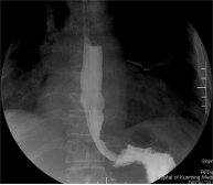
Figure 1. Upper Gastrointestinal Imaging: Lower Esophageal Stenosis, Filling Defect, Passable Contrast, Possible Esophageal Occupancy.
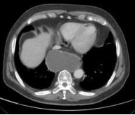
Figure 2. Chest CT Enhancement Scan: Cystic Liquid Hypodense Shadow is Seen in the Lower Esophageal Region, Which is Closely Related to the Esophagus, with the Possibility of Submucosal Encapsulated Effusion or Parietal Cyst of the Esophagus.
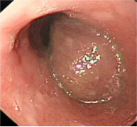
Figure 3. Gastroscopy: a Bulge was Seen in the Esophagus 32 cm from the Incisors, with Narrowing of the Esophageal Lumen and External compressive Stenosis.
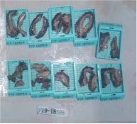
Figure 4. Postoperative Gross Pathology: the Eye Saw Multiple Blocks of Grayish-White Brown Tough Cystic Wall-Like Tissue with a Wall Thickness of about 0.5-1 cm and Rough Inner Wall of the Capsule.